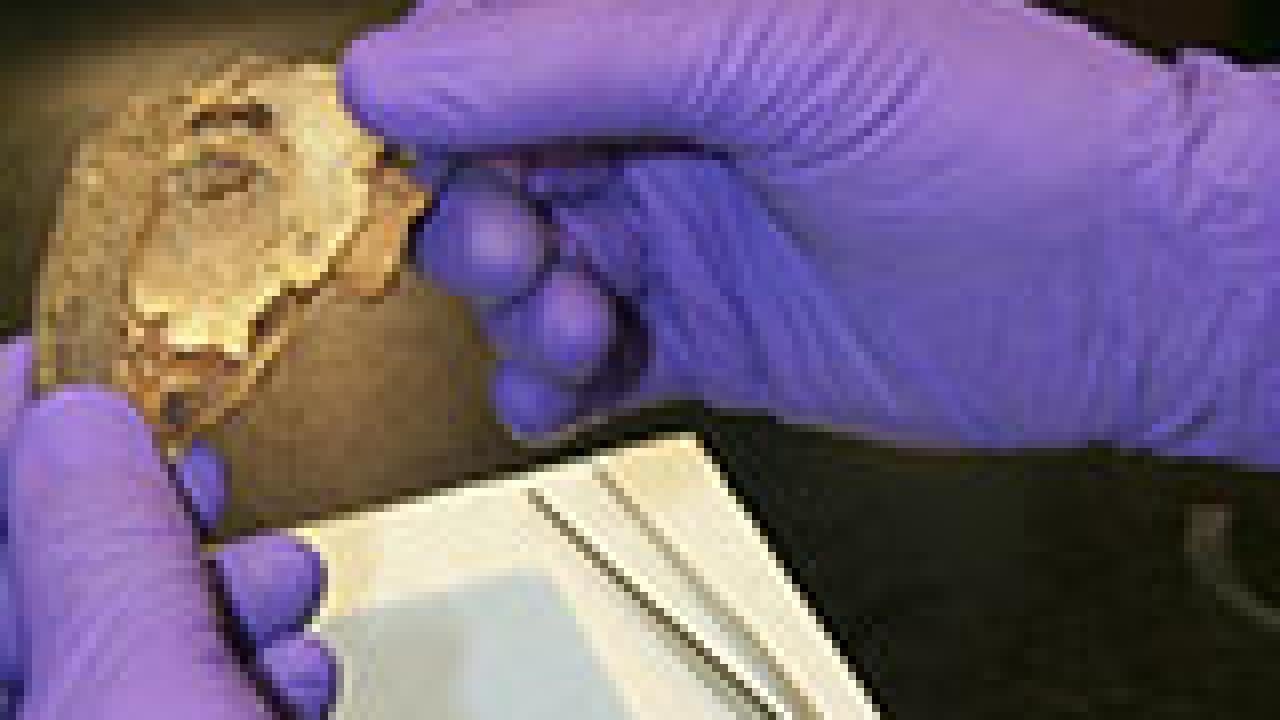
During the past week, ophthalmologist William Lloyd has been dissecting and examining the eyes of two North Chilean mummies for evidence of various diseases and medical conditions.
One of the eyes belonged to a boy who was 2 years old when he died 1,000 years ago, and the other is from a woman, who was approximately 23 years old when she died 750 years ago.
"The opportunity to analyze two pre-Columbian era mummy eyes is exciting and fascinating," said Lloyd, an accomplished physician, researcher, professor, author and expert in comparative ophthalmology, which involves the study of the eye across species. Lloyd holds joint appointments in the departments of Ophthalmology and Vision Science, and Pathology at the UC Davis School of Medicine.
"By analyzing these eyes," Lloyd continued, "we hoped to determine if their pathology suggests any so-called modern day diseases, like diabetes or high blood pressure. And while there was no immediate evidence of such diseases, we'd like to do more research."
The project began when Huck Holz, chief resident in the Department of Ophthalmology and Vision Science, read an article about the founder of modern paleopathology, Arthur Aufderheide, in the May 16 issue of the New Yorker magazine. Paleopathology, the study of ancient diseases, has taken Aufderheide around the globe, salvaging mummies' organs and tissues in various stages of decomposition.
The thin tissues that make up the eye allow it to dehydrate quickly and, because moisture causes decay, most mummies are found with well-preserved eyes.
In the New Yorker article, Aufderheide said that he has been saving the eyes for the right investigator, someone with the expertise and the commitment to examine them thoroughly. Holz and Lloyd convinced Aufderheide that they were the researchers he had been waiting for.
Microscopic viewing
Examining the specimens involved rehydrating the eyes and optical nerves, preparing the tissues for chemical processing, embedding the tissues in paraffin, slicing the specimens for microscopic viewing, applying stains to highlight selected cellular characteristics, and finally examining the tissues under a microscope.
Cheers and applause erupted when the stained specimen of the 23-year-old female was slid onto the microscope's stage and the well-preserved tissues appeared on the monitor.
"We were hoping to see recognizable tissue, but this is better than any of us imagined," said Lloyd. "This tissue looks just like it came from someone who had eye surgery yesterday."
The other mummy's eye did not fare as well. The tissue was decomposed from a millennium of exposure to the elements.
Initial tests showed no obvious signs of eye diseases, such as glaucoma or macular degeneration, but more tests are necessary to be conclusive. Lloyd hopes to examine more mummy eyes and says there are many systemic ailments that can be found by examining the eyes.
"During modern-day eye exams, we can see signs of diabetes, high blood pressure, various cancers, nutritional deficiencies, fetal alcohol syndrome and even early signs of HIV infection," said Lloyd. "We hope to find something new about what kind of ailments might have afflicted ancient people."
Pneumonia cited
Both mummies in this study were already known to have recovered from pneumonia. One of the woman's lungs was adherent to her chest wall and both of the young boy's lungs were adherent to his chest wall.
"This adherence is consistent with a recovery from pneumonia," said Aufderheide, who is a professor of pathology at the University of Minnesota, Duluth, School of Medicine. "Since we see it on both of the boy's lungs, he probably had and recovered from pneumonia, twice."
The child, who was one of the last members of the Tihuanacu culture, also had an inherited cystic disease in his liver.
"We're not sure if the liver disease is what killed him," said Aufderheide. "There were a few preserved internal organs, but most of the body was in decay."
Future studies possible
The 23-year-old woman was buried in a seated position, fully clothed in embroidered V-neck wool shirts. She wore sea-lion-hide sandals and, on her head, a bandana. Her hair was in two braids. In addition to the pneumonia, she had lice, bad teeth and osteoporosis.
"It's likely that the young woman's osteoporosis was caused by a diet that included oxalate-producing plants, which inhibits the body's ability to assimilate calcium," said Aufderheide.
Perhaps future studies will reveal more information about the lives and deaths of these two people and their contemporaries.
Kelly Gastman is a senior information officer for the UC Davis Health System.
Media Resources
Clifton B. Parker, Dateline, (530) 752-1932, cparker@ucdavis.edu