Technology is aiding research in autism, meditation and beyond
UC Davis brain researchers are exploring how kids with autism do math and what happens when Buddhist monks meditate.
Functional magnetic resonance imaging, or fMRI, lets scientists at UC Davis' Center for Mind and Brain see precisely which areas of the brain are at work during different tasks.
The imaging technique is the younger sibling of magnetic resonance imaging, or MRI, a noninvasive tool that revolutionized radiology and earned its inventors the 2003 Nobel Prize in medicine. "Functional magnetic resonance imaging allows us to look at the normal, functioning human brain," says Ron Mangun, director of the Center for Mind and Brain.
Susan Rivera, a new colleague of Mangun's in the UC Davis psychology department, uses functional MRI for her research on autism. Instead of describing unusual behaviors in children with autism, Rivera's research will show the autistic brain at work. She conducts her studies at the UC Davis Medical Investigation of Neuro-developmental Disorders (M.I.N.D.) Institute, one of the nation's leading centers for autism research.
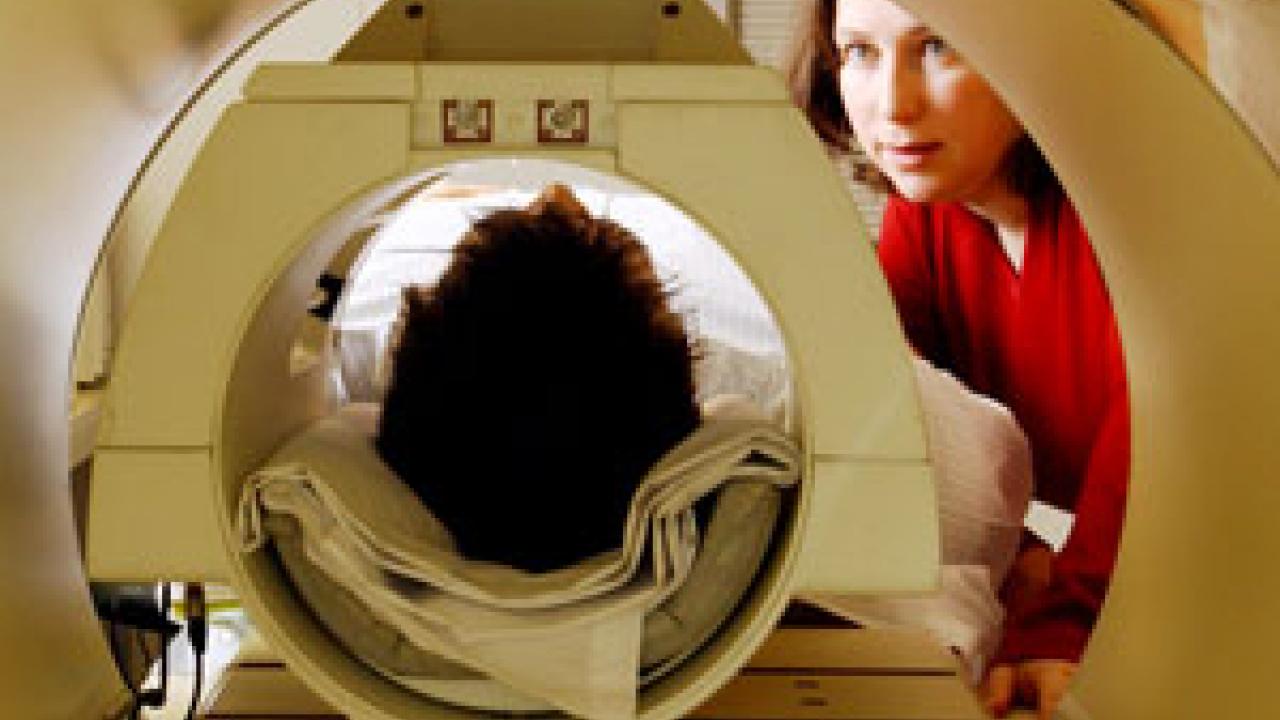
During an ordinary MRI scan, the patient lies inside a very large magnet. In this strong magnetic field, radio-frequency pulses elicit tiny signals from the body's hydrogen atoms. The signals are mathematically transformed into a precise map of soft tissue, which is used to diagnose everything from tendonitis to brain tumors.
The newer technology goes further, letting scientists visualize brain function. In Rivera's studies, a child with autism lies inside a scanner equipped with a viewing screen and a response button. This allows Rivera to give the child specific tasks. While his brain works on the tasks, the scanner measures the magnetic properties of his hemoglobin, the oxygen-carrying protein in his blood. Hemoglobin's magnetic signal changes when oxygen binds to it. The scanner exploits this change to show precisely where oxygen-rich blood is flowing in the child's brain -- and more oxygen-rich blood means more nerve activity.
To study brain wiring in autism, Rivera scans children with autism performing tasks they do well. "Scientists usually study adults with autism doing things that they're really bad at, like assessing emotions," she says. But those studies give an incomplete picture of the autistic brain. For example, many people with autism excel at math, but no one knows how their brains attack math problems. "Autistic kids may be using their brains in a really different way" than typically developing children, Rivera says. Figuring that out might open new doors in autism treatment.
Detailed images from functional scans also help Ron Mangun and colleague Charan Ranganath study attention and memory. They hope to shed light on problems such as attention deficit disorder and memory problems in the elderly.
Mangun examines selective visual attention: the ability to filter important bits of information from everything we see. Without this focus, he says, ordinary tasks like driving a car would be impossible. He's exploring how the attention filter works. He wonders what happens between the eye, which takes in all visual signals, and the mind, which knows that some sights matter more than others.
Before fMRI, neurologists attacked these questions by looking at behavioral changes in brain-injured patients. "We would infer something about the normal brain based on the broken brain," Mangun says. But that approach can be misleading. Functional brain damage spreads far beyond the injured area, which made behavioral measurements poor tools for understanding the effects of an injury.
Now, functional scans show the workings of the whole, healthy brain at once. This lets Mangun identify precisely which areas of the brain take charge of different kinds of visual attention, and measure how hard each area works in response to certain cues. He has found, for example, that certain nerve networks connecting the brain's frontal lobe, behind the forehead, and the parietal lobe, at the top of the head, are specialized to pay attention to objects' locations. Such data lets scientists puzzle out how the brain coordinates selective visual attention. This is the first step towards helping people with disordered visual attention -- which Mangun says could include everyone from garden-variety klutzes to those with Attention Deficit Disorder or schizophrenia.
The technology makes sense of puzzling behavioral data in other situations, too. Ranganath, who conducts research with the UC Davis Center for Neuroscience, uses it to study memory problems in older people. "We can use this technology to relate brain function questions to questions about brain anatomy, uncovering connections between the mind and the brain," he says.
The elderly people Ranganath studies have "mild cognitive impairment." For the most part, their brains work normally, but they have some memory problems and are at increased risk for dementia and Alzheimer's disease.
By performing fMRI scans while they did different memory tasks, Ranganath and his colleagues were able to put their subjects in two categories. Some had defects in the brain's white matter, which impaired their working memory. "Working memory is what you're keeping active in the brain," Ranganath explains. Working memory holds small sets of information for short periods of time. Others showed atrophy, or shrinking, of the hippocampus. They experienced defects in episodic memory, the memory of specific events. Understanding these sub-types of mild cognitive impairment could help scientists develop better treatments for memory problems in the elderly.
The researchers at the Center for Mind and Brain collaborate with scientists within and outside UC Davis. For example, they work with both the UC Davis M.I.N.D. Institute and the UC Davis Research Imaging Center at the UC Davis Medical Center.
Mangun also has a more unusual collaboration -- with a group of Buddhist monks. This project will ask whether people can learn to focus their visual attention more effectively. Monks claim that, during meditation, they can hold a single visual image in their minds for unusually long periods of time. To see if learning Buddhist meditation techniques enhances the brain's ability to focus, Mangun's team will measure selective visual attention in people before and after intensive training in Buddhist meditation. "We know almost nothing about the plasticity of attention systems," Mangun said. "What if there could be huge plastic changes in these systems? Could we train people out of attention disorders?"
He thinks functional MRI makes answers to these questions more accessible than ever before. He says that "this tool will lead us to the 'big questions' about awareness and consciousness."